4 Unique Uses Of Raman Spectroscopy

By John Oncea, Editor

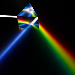
Spectroscopy began when Isaac Newton split light with a prism and today is used in countless ways in medicine, physics, chemistry, and astronomy. Here, we look at Raman spectroscopy and how it is being used in new and unique ways.
There are, by my count, at least 250 materials analysis methods. Of those, depending on the source, there are 12, five, 22, or eight, or (insert your favorite number here*) types of spectroscopy. To keep this article at fewer than 10,000 words, let’s focus on one type of spectroscopy, Raman spectroscopy, and some unique ways it’s being used.
To make sure we’re all on the same page here, let’s define some of the terms we’ll be discussing.
- Materials analysis method: Identifying the chemical and structural composition of a material to understand its derived properties.
- Spectroscopy: The branch of science concerned with the investigation and measurement of spectra produced when matter interacts with or emits electromagnetic radiation.
- Raman spectroscopy: A spectroscopic technique typically used to determine vibrational modes of molecules, although rotational and other low-frequency modes of systems may also be observed. Raman spectroscopy is commonly used in chemistry to provide a structural fingerprint by which molecules can be identified.
Put it all together and we’ve got a technology that plays a crucial role in the realm of graphene research, providing valuable information regarding the number of layers present, their orientation, and the quality and types of edges. By subjecting graphene samples to various perturbations such as electric and magnetic fields, strain, doping, disorder, and functional groups, Raman spectroscopy can reveal how these perturbations affect the material's properties.
Moreover, this analytical technique is not limited only to graphene but also can provide insights into all sp2-bonded carbon allotropes, as graphene is their fundamental building block. Its ability to capture rich details and facts has made Raman spectroscopy an indispensable tool in the field of carbon-based materials.
* Mine is 25 – the number I was given when I made my elementary school basketball team in the fourth grade.
Cell Cultures, Bacteria Identification, Blood Detection, And Pharmaceutical Analysis
Now that we’re all on the same page as to what Raman spectroscopy is, let’s take a look at four unique ways it is being used starting with real-time monitoring of cell cultures.
A team of researchers from Kwansei Gakuin University’s School of Biological and Environmental Sciences and Yokogawa Electric Corporation collaborated to develop a novel Raman calibration model for real-time monitoring of cell cultures, reports Spectroscopy Online.
“Real-time analysis of bioprocess parameters is essential to increase efficiency and reduce production costs’” writes Spectroscopy Online. “Currently, some parameters, such as metabolite and product concentrations, cell growth, and other quality attributes, are monitored using offline methods.
“According to the research team of Risa Hara, Wataru Kobayashi, and Yukihiro Ozaki, this new method uses a conventional Raman spectroscopy technique of analyzing each component in the culture media while offering the ability to calibrate multiple components in real-time. This allows researchers to better control the cell culture environment and improve culture efficiency.”
The method involved detecting the specific peak positions and intensities for each component and preparing the samples by mixing them as per the experimental design. The Raman spectra of these samples were analyzed to build the calibration models, and several combinations of spectral pretreatments and wavenumber regions were compared to optimize the model for cell culture monitoring, without using the culture data.
The model's accuracy was evaluated by conducting actual cell culture activities and comparing the in-line measured spectra with the developed calibration model. The results showed that the calibration model is highly accurate for three essential components, namely glucose, lactate, and antibody. The root mean square errors of prediction (RMSEP) for glucose, lactate, and antibody are 0.23, 0.29, and 0.20 g/L, respectively. This novel method enables the real-time measurement of vital parameters like metabolite and product concentrations, cell growth, and other quality attributes related to products, without using offline methods.
“Real-time monitoring and control of bioprocess parameters are essential to increase efficiency and reduce production costs,” Spectroscopy Online writes. “The new Raman calibration model offers a noninvasive method for analyzing components in culture media and enables the calibration of multiple components in real-time, which allows for better control of the culture environment and improves culture efficiency. This method has presented innovative results in developing a culture monitoring method without using culture data, whereas using a basic conventional method of investigating Raman spectra. It is expected that this new method will be put into practical use and become more widely used in the future.”
Next up is the use of Raman spectroscopy, combined with acoustic bioprinting and machine learning, as a novel technique for rapid bacterial identification. This innovative technique promises improved clinical diagnosis, safer food, faster drug development, and enhanced environmental monitoring, according to AZoOptics.
Currently, diagnosing bacterial infections requires culturing samples and prescribing broad-spectrum antibiotics while waiting for results which can take several days. This leads to nearly one in three patients receiving unnecessary treatment and contributes to developing antimicrobial resistance.
But the label-free, non-invasive use of Raman spectroscopy to identify bacterial species takes advantage of the unique molecular structure of each type and cell strain, giving rise to a distinct spectral fingerprint that can be used for identification. “By illuminating a sample with laser light, Raman spectroscopy measures the energy scattered (weak Raman scattering) during interactions between the light and the sample molecules,” writes AZoOptics. “Then, it filters out the laser light to analyze the captured signal and match it with known bacteria.”
Raman spectroscopy offers numerous benefits when compared to other forms of tests used for bacterial identification, such as nucleic-acid-based polymerase chain reaction or protein-based enzyme-linked immunoassay. One of the most significant advantages of Raman spectroscopy is that it utilizes low amounts of reagents and requires minimal sample preparation. Additionally, the equipment used for this process is low-cost, and the analysis is non-destructive, adding to the cost-efficiency of the process.
But the big win comes when combing it with acoustic bioprinting and machine learning. Stanford University researchers “used the acoustic droplet ejection (ADE) technique to isolate the cells in extremely small samples and to eliminate unwanted spectral information,” AZoOptics writes. “ADE uses ultrasonic waves that create radiation pressure to eject a droplet from the surface, which is only a few dozen-sized cells.
“The team added gold nanorods to the samples, which bind to bacteria and intensified the Raman signal 1,500 times. They then employed machine learning to compare the different light patterns emitted by each printed fluid dot to identify the unique Raman spectral signatures of any bacteria in the sample.”
The proposed methodology not only offers a remarkable level of throughput but also presents a tremendous potential for speedy and cost-effective on-site diagnosis without the need for laboratory analysis. This feature makes it appropriate for personnel with minimal training. Additionally, the technique promises to provide faster, more precise, and inexpensive microbial assays for a diverse range of fluids when compared to traditional culturing methods, which can take several hours or even days. These benefits could be game-changing on the front lines of medical care, where rapid and accurate diagnoses can often mean the difference between life and death. With the ability to rapidly diagnose and treat numerous infectious diseases cost-effectively, this technique could significantly improve public health outcomes on a global scale.
Third on our list is a novel Raman spectroscopic method for detecting traces of blood on an interfering substrate as reported by Nature. Guys, I’m not going to pull any punches here. This is a long, technical article that would take many, many words to sum up. That said, I’m going to drop the text from the abstract here and you can click the link if you want to get deeper into the weeds.
“Traces of body fluids discovered at a crime scene are a primary source of DNA evidence. Raman spectroscopy is a promising universal technique for identifying biological stains for forensic purposes. The advantages of this method include the ability to work with trace amounts, high chemical specificity, no need for sample preparation, and a non-destructive nature. However, common substrate interference limits the practical application of this novel technology. To overcome this limitation, two approaches called "Reducing a spectrum complexity" (RSC) and "Multivariate curve resolution combined with the additions method" (MCRAD) were investigated for detecting bloodstains on several common substrates. In the latter approach, the experimental spectra were “titrated” numerically with a known spectrum of a targeted component. The advantages and disadvantages of both methods for practical forensics were evaluated. In addition, a hierarchical approach to reduce the possibility of false positives was suggested.”
Finally, Spectroscopy Online is back with a look at Raman spectroscopy and its applications in pharmaceutical analysis. “One of the significant advantages of Raman spectroscopy is its insensitivity to water molecules,” writes Spectroscopy Online. “This attribute makes the technique superior to other methods in detecting various drugs in drug delivery systems. Additionally, Raman spectroscopy allows for the rapid measurement of vibrational energy states in a contactless and non-destructive manner, making it an attractive technique for biomedical and pharmaceutical analysis.”
The versatility of Raman spectroscopy permits its use in detecting counterfeit drugs, as well as for the analysis of the interactions between drugs and polymers. In addition to its applications in traditional pharmaceutical analysis, Raman spectroscopy also has demonstrated potential in the analysis of herbal medicines and illicit drugs.
Furthermore, Raman spectroscopy has several advantages over other analytical methods in the field: It requires little or no sample preparation, and it can identify and quantify multiple components simultaneously.
Raman spectroscopy is a non-destructive and non-invasive method of analysis. It does not require a change to the sample being analyzed, which is extremely beneficial when dealing with expensive or difficult-to-obtain samples.
