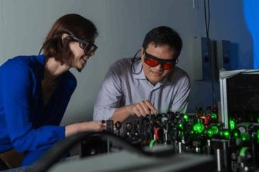
STReM Technique Captures High-Speed, Super-Resolution Images Without Need For A Fast Camera
By Jof Enriquez,
Follow me on Twitter @jofenriq

Rice University researchers have refined a Nobel Prize-winning microscopy technique to capture fluorescing molecules at a frame rate 20 times faster than other laboratory cameras.
The 2014 Nobel Prize in Chemistry was awarded for the development of super-resolved fluorescence microscopy, which allowed viewing of images in 250-nanometer resolution — smaller than half the wavelength of light. Despite the unprecedented high-spatial resolution achieved, the temporal resolution remains low, meaning one still can't take pictures of anything faster than the frame rate, which maxes out at 10 to 100 milliseconds for a typical charge-coupled device (CCD) camera.
Researchers at Rice University say they have overcome this limitation by developing a technique called super temporal resolution microscopy (STReM), which improves temporal 2D super-resolution microscopy by a factor of 20, and captures biological processes happening at 10 to 100 frames per millisecond — such as protein absorption and detailed molecular motion.
"This is achieved by rotating a phase mask in the Fourier plane during data acquisition and then recovering the temporal information by fitting the point spread function (PSF) orientations," the researchers wrote in The Journal of Physical Chemistry Letters.
Point spread functions are a measure of the shape of images both in and out of focus. STReM uses point spread function changes from the spinning mask to collect temporal information.
"As the mask spins faster than the camera can shoot, the image sensor picks up the molecule’s movement — so one molecule pops up several times in the same image. Since the phase mask allows a trained eye to determine distance, researchers can then measure the copies of the molecule on the image and see where and how it moved," explains Digital Trends.
Since the STReM technique compresses faster information in a slower camera frame rate, labs don't necessarily need faster cameras to capture super-resolution images. The Rice researchers claim to have only spent a few hundred dollars designing and building the mechanism. Moreover, STReM is ideal for imaging delicate objects like biomolecules, which can be damaged under powerful electron microscopes.
“The purpose is to allow scientists to study fast processes without the need to buy faster and much more expensive cameras,” said Rice graduate student Wenxiao Wang, lead author of the paper, in a news release. “This involves extracting more information from single images.”
Another new technique to sharpen images captured through nanoscale microscopy (nanoscopy) was developed by scientists at the Joint Quantum Institute (JQI). They described a procedure to correct an unwanted image-dipole effect to produce better super-resolution images.
