Smartphone Microscope Detects, Measures Individual DNA Molecules
By Chuck Seegert, Ph.D.

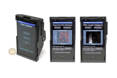
Researchers from UCLA’s California NanoSystems Institute have imaged and measured the sizes of individual DNA strands using a smartphone. Their newly developed, lightweight, and compact device cheaply transforms an off-the-shelf smartphone into a fluorescent microscope with advanced capabilities.
Fluorescent microscopes are often used to diagnose cancer and study neurodegenerative diseases like Alzheimer’s. After samples are labeled with fluorescent molecules, a laser is used to excite the molecules. The microscope can then detect the light emitted by the labeled molecules. This method is routinely used to study objects that are several orders of magnitude smaller than the diameter of a human hair. Generally, however, these instruments are bulky, expensive, and complicated to operate, which confines them to high-tech laboratories staffed with highly trained personnel.
In an effort to make this technology available to more researchers, a team from UCLA has simplified and streamlined the process, according to a recent article from the UCLA Newsroom. The unit is portable, inexpensive, and is based on a 3D printed optical device that attaches to a smartphone. Once attached, it uses the phone’s camera while creating a high-contrast, dark-field imaging setup. An inexpensive lens uses thin-film interference filters and a laser diode to fluorescently excite the labeled DNA molecules. The DNA is studied on a miniature dovetail stage and can be visualized and measured for length.
Though the device images the DNA molecule, analysis is undertaken via a computational framework that interfaces with the phone through a custom app, according to a recent study published by the team in ACS Nano. Length measurements of the individual DNA molecules are measured on a remote server, and the data is returned to the smartphone after analysis is complete. So far, the sizing accuracy of the system is <1 kilobase-pairs (kbp) for DNA specimens of 10 kbp or longer, and the field-of-view is about 2 mm2.
The new system has been developed under the direction of Aydogan Ozcan, the Chancellor’s Professor of electrical engineering and bioengineering at the UCLA Henry Samueli School of Engineering and Applied Science, according to the press release. A focus of the team’s research is translating microscopy and sensing techniques to field-portable, cost-effective instruments that could allow high throughput studies to be performed. The team’s efforts are centered on positively impacting research and educational efforts in resource-limited countries.
Similar efforts are underway elsewhere to enable diagnostic laboratory services in third-world countries. For example, a recent story on Med Device Online described a Harvard engineering team’s efforts to develop a handheld device that performed many different clinical assays and transmitted results via a cell phone.
Image Credit: Ozcan Lab at UCLA
