Optical Imaging Through Opaque Layers
By Chuck Seegert, Ph.D.

If you were to shine a light on someone’s skin, you wouldn’t expect to see what’s going on behind it. With a new technique published in The Optical Society’s journal Optica, however, you can do just that with surprisingly high levels of resolution.
“Imagine shining a flashlight through a thick fogbank to try to see a single dot,” said Yaron Silberberg, a researcher at the Weizmann Institute of Science in Israel, in a recent press release. “The light would become so scattered as it traveled through the fog that you wouldn’t be able to make out what was hidden inside. By carefully shaping the light going in, however, it would be possible to home in on your target. That is exactly what the researchers were able to achieve in a way no one has ever done before.”
Similar results have been achieved historically, but only with the use of a reference point called a “guide star,” according to the press release. A guide star is a term developed in astronomy. Essentially, it is a light source in the same visual field as the one you are studying that you can use as a baseline data point. Seeing how light from the guide star is behaving when it encounters something like the atmosphere can eliminate “twinkling” and increase the resolution of the dimmer stars under study.
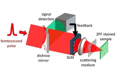
In medicine and biology, though, placement of a guide star object, like a fluorescent particle, is generally invasive and can disturb surroundings and damage tissue. The newly developed approach, however, takes feedback from the surrounding tissue and adjusts the light going into the sample to compensate for scattering. This advance eliminates the need for the invasive guide star.
“What we have discovered is that it’s possible to efficiently 'pre-correct' the laser beam using the nonlinear fluorescence signal,” noted Ori Katz, a scientist at the Langevin Institute in Paris, France, and co-author of the study. “The end result is that instead of having a distorted, blurred light source on the object to be imaged, we have a tightly focused, or in this case, refocused beam of light.”
This technology uses femtosecond laser pulses and can image objects with resolutions on the single digit micron scale. Details of the team’s research are published in a study in the journal Optica.
The non-invasive study of tissues is a topic of research that continues to accelerate. Recently in an article on Med Device Online, another non-invasive, optical method was discussed that identifies different characteristics of skin cancer.
Image Credit: Optica
