New Photoacoustic Device Provides Non-Invasive, Deep-Tissue Imaging
By Chuck Seegert, Ph.D.

Until now, there has been no suitable method to measure the depths of tumors and create maps of how extensive those tumors are. Researchers from Washington University in St. Louis, however, have developed a new device that uses photoacoustics in order to address that problem.
Some of the most prevalent forms of cancer are those found in the skin. Of these, Melanoma is the most deadly — accounting for up to 75 percent of skin cancer-related deaths in the U.S. A key to understanding the aggressiveness of Melanoma is to determine its thickness, something that is currently a challenge even with a biopsy.
Techniques using light and other spectroscopic energies are being developed to non-invasively study skin cancers, but they are often limited to the surfaces of tumors. When light hits skin, it tends to scatter, which prevents it from penetrating to the appropriate depth of about 2 to 4 millimeters. More traditional imaging methods like high-frequency ultrasound and MRI are expensive and generally don’t have the resolution or contrast necessary to delineate the extent of the tumor.
"Any type of tissue diagnosis at this point in time requires taking tissue out of the person and processing it and looking at it under the microscope," said Dr. Lynn Cornelius in recent press release from The Optical Society (OSA). Cornelius is a coauthor of the study that described the new device.
While traditional diagnostics will give information on the sample taken from a small area of the tumor, the whole volume of the tumor can’t be understood without taking multiple biopsies. Often these tumors are in cosmetically important areas like the face, so minimizing the biopsies taken is often attempted. Without a proper understanding of the tumor’s extent, however, surgery can be less effective. If the tumor is deeper than expected, it may even lead to a second operation.
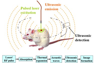
To overcome these limitations, the research team developed an instrument that uses a phenomenon called the photoacoustic effect. Photoacoustics is the transformation of light energy into vibrations. These vibrations, in turn, can travel through tissue more reliably than light.
As a laser beam shines onto the surface of the skin, it interacts with melanin, a pigment contained in the skin. Melanin absorbs the light and transfers it into high frequency acoustic waves. Because tumors contain more melanin than surrounding tissues, the boundaries can be identified and used to map the entire tumor in a high resolution, 3D image.
In a research study published in the OSA’s Optics Letters, the latest prototypes of the new device have been used to generate images of tumors and their underlying surfaces, providing very accurate measurements of their depths. This was demonstrated in artificial model tumors, as well as in vivo in a mouse tumor model.
Currently the photoacoustic imaging system is being used in clinical trials, and the team is hopeful that it will be commercialized.
Portable diagnostics are an area of medicine that is currently receiving intense focus. For example, recently laser technology and fiber optics were combined to diagnostically study melanoma tumors in a similar effort to more accurately diagnose tumors without performing extensive biopsies.
Image Credit: “PASchematics v2.” Bme591wikiproject at the English language Wikipedia. 2007 CC BY-SA 3.0: http://creativecommons.org/licenses/by-sa/3.0/
