Minimally-Invasive Brain Imaging May Be Possible With Surgical Needles

Scientists have developed a novel brain imaging technique that blends existing methods, using lasers and endoscopes to study brain tissue. By substituting a glass surgical needle for the endoscope, a proof-of-concept study in mice has demonstrated technology that might lead to a minimally invasive method for imaging deep brain tissue, one that could provide a better understanding of neurological conditions.
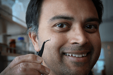
Rajesh Menon and his team at the University of Utah have been studying the potential of computational cannula microscopy (CCM) for deep tissue imaging. In a study published in Applied Physics 2015, the Utah scientists first unveiled their idea to convert an ordinary glass needle — or cannula — into a high-resolution microscope, which could overcome obstacles presented by currently available technologies.
“Multi-photon microscopy (MPM) can achieve [brain tissue imaging], but at limited depths and at limited resolution,” wrote the researchers in their most recent study, published by Scientific Reports. Imaging through MPM, explained the scientists, was only possible by imaging tissue within 1.2 mm of the brain’s surface, and previous attempts to refine the method through the use of longer wavelengths were stymied by poor resolution caused by scattered light. Alternatively, brain imaging can be accomplished via endoscope, but the scopes’ size makes the procedure likely to cause brain trauma.
“What we have done is to take a surgical needle that’s really tiny and easily put it into the brain as deep as we want and see very clear high-resolution images,” said Menon to UNews. “This technique is particularly useful for looking deep inside the brain where other techniques fail.”
Menon and his team — which included Nobel Prize-winning scientist Mario Capecchi and lead author Ganghun Kim — used their needle microscope to shine laser light “like a flashlight” into the brains of mice, which had been engineered so that cells the scientists wanted to view were glowing. The needle then collected the light from that glow using a standard camera, and the data was run through an advanced algorithm that reassembles the scattered light into an image.
According to Menon, this technology has proven effective with mice and could prove useful in humans for imaging the brain and other organs. For now, Menon intends to continue studying mouse brains for any insight they can provide into neuro mechanics and conditions.
“Our motivation for this project right now is to look inside the brain of the mouse and further develop the technique to understand fundamental neuroscience in the mouse brain,” Menon told UNews.
Recent research from the University of Adelaide has likewise repurposed surgical needles to provide imaging, and has developed a system of “smart needles” that could guide surgeons through complicated brain surgeries. The system uses fiber optic camera and infrared light to image pathways through the brain, and advanced computer software is programmed to recognize blood vessels and send alerts to surgeons, preventing a potentially dangerous bleed.
