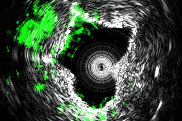
Laser-Enabled, Label-Free Imaging Provides 3D Look Inside Arteries
By Chuck Seegert, Ph.D.

A new medical imaging system that produces 3D images of the insides of arteries has recently been developed by researchers at Purdue University. The minimally invasive equipment uses a fast-pulsing laser that heats and expands tissues, generating measurable ultrasound frequencies and enabling the identification of dangerous plaques.
Plaque buildup in the coronary arteries is a common cause of heart disease. The waxy substance can accumulate over time and begin to block blood flow through the vessels, a condition known as atherosclerosis. If left unattended, plaque will eventually harden, which predisposes it to crack or rupture. Ruptures of plaque can cause clots in the vessel and close it off completely, causing a heart attack.
To diagnose the plaque more accurately, a team at Purdue University has improved upon a technique called “intravascular photoacoustic imaging” (IVPA), according to a recent press release from the university. The system uses a pulsed laser that cycles at up to 2,000 pulses per second, revealing the carbon-hydrogen bonds that compose the lipid molecules in the plaque.
"This allows us to see the exact nature of plaque formation in the walls of arteries so we can define whether plaque is going to rupture," said Michael Sturek, a professor and chair of the Department of Cellular & Integrative Physiology at Indiana University School of Medicine, according to the press release. "Some plaques are more dangerous than others, but one needs to know the chemical makeup of the blood vessel wall to determine which ones are at risk of rupturing."
To date, attempts at making IVPA a viable clinical solution have been hampered by slow imaging speeds, according to a recent study published by the team in Nature Scientific Reports. Previous systems have only been capable of generating an image once every 50 seconds, but the new system has reduced it to 1.0 seconds per frame. The improvement was made possible by a custom 2-kHz master oscillator power amplifier-pumped, barium nitrite Raman laser.
"This innovation represents a big step toward advancing this technology to the clinical setting," said Ji-Xin, a professor in the Weldon School of Biomedical Engineering and the Department of Chemistry at Purdue University, according to the press release.
The rapidly pulsing laser heats and expands the tissue. While the heat isn’t enough to damage tissues, the fluctuation is enough to be detected as an ultrasound signal by a sensor in the vicinity, according to the press release. Additionally, this imaging technique does not require labels, which makes it more attractive for diagnostic applications.
Predicting risk of heart attack and stroke is an important diagnostic tool that could save many lives. Recently in an article on Med Device Online, another diagnostic approach using MRI was discussed for identifying heart attack and stroke risks in diabetics.
Image Credit: Purdue University Weldon School of Biomedical Engineering image/Ji-Xin Cheng
