How Optics And Flow Cytometry Are Improving Pancreatic Cancer Treatment

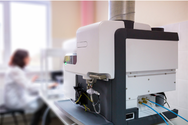
Researchers at the University of Texas at Austin are advancing cancer treatment by using an optical system, the mechano-chemical flow cytometer (MCFC), to analyze the mechanical and chemical properties of cancer cells. This innovative system employs flow cytometry and spectroscopy, specifically broadband coherent anti-Stokes Raman spectroscopy (BCARS) and real-time deformability cytometry (RT-DC). Together, these techniques provide a label-free way to assess cancer cell characteristics, which can reveal the invasiveness and treatment resistance of cancers, especially aggressive types like pancreatic cancer. Unlike traditional fluorescent-tagging methods, this approach avoids cell damage from tagging, thus preserving critical information about both the surface and internal structures of cells.
The MCFC combines advanced imaging technologies to better understand cancer cell behavior and refine therapeutic approaches, potentially improving survival rates. This device enables precise predictions of how specific cancers will respond to different treatments, allowing clinicians to tailor approaches more effectively. The Parekh Lab, which developed the MCFC, received the 2020 Edmund Optics Silver Educational Award, supporting its efforts to bring this technology closer to clinical application. Once commercially available, the MCFC could be transformative in providing individualized, data-driven cancer treatments, particularly for patients with hard-to-treat cancers.
Get unlimited access to:
Enter your credentials below to log in. Not yet a member of Photonics Online? Subscribe today.
