How Optical Coherence Tomography Is Helping Shape The Future Of Photonics

By John Oncea, Editor

OCT is a noninvasive imaging method that uses reflected light to create pictures of the back of your eye. It helps eye care providers diagnose and manage common eye diseases, and its popularity is helping shape the future of photonics.
Technology evolves for any number of reasons. Sometimes, it’s as a result of a human need, such as COVID-19 accelerating mRNA vaccine development, solving an urgent health crisis and reshaping biotech R&D in the process.
There are also economic incentives. For instance, the rapid advancements made to smartphones by Apple, Samsung, and others are, in large part, happening to meet consumer demand for more speed and better cameras (as well as to make a buck or two).
Competition amongst nations is a major driver of technological change. If it weren’t for the space race between the U.S. and USSR, there might never have been any memory foam mattresses, scratch-resistant lenses, cordless tools, wireless headsets, athletic shoes, invisible braces, CAT scans, smoke detectors, or cochlear implants. *
Then there are innovations that emerged as unexpected discoveries during experiments – happy little accidents, if you will. Take Post-it Notes, for example. These ubiquitous pieces of paper used for leaving notes, reminders, or messages came about when Spencer Silver, a 3M scientist, was trying to create a strong adhesive but instead developed a weak, reusable adhesive. This “failed” adhesive later became the basis for the Post-it Note, initially intended to hold bookmarks in place.
The first version of Coca-Cola was a failed attempt by John Pemberton to create a non-alcoholic alternative to wine-based drinks, and it occurred when he accidentally mixed syrup with carbonated water. And almost everyone knows that penicillin was the result of bacteriologist Alexander Fleming returning from a vacation, only to discover a mold, Penicillium notatum, had contaminated a petri dish of bacteria and prevented the bacteria from growing around it. Almost 100 years later, Fleming’s happy little accident has prevented millions of deaths from infection and disease.
Which brings us to optical coherence tomography (OCT), a non-invasive imaging technique that uses light waves to create detailed, cross-sectional images of the eye’s structures, whose surge in popularity is catalyzing a wave of innovation, helping shape the future of photonics.
* There would be Tang, Velcro, and Teflon, however. Contrary to popular belief, NASA did not invent these things.
What Is Optical Coherence Tomography
Optical coherence tomography, or OCT, is a noninvasive imaging method that produces cross-sectional images of the eye. According to the Cleveland Clinic, the technology measures wavelengths of infrared light that reflect off the retina, creating layered images that allow specialists to measure the depth and structure of critical tissues. This level of detail helps in diagnosing and managing a range of eye conditions.
When OCT is used to examine the eye, it is sometimes called ocular coherence tomography. In recent years, its use has expanded beyond ophthalmology, and specialists in cardiology, neurology, and oncology have adopted OCT to examine blood vessels and other tissues. For example, OCT angiography provides a new way to visualize vascular networks without the need for dye injections.
A provider may suggest OCT if symptoms or exam findings point to certain eye conditions. It may also be recommended for people at higher risk of age-related eye problems or other eye diseases. Because OCT can be repeated over time, comparing scans can help detect even subtle changes and guide decisions about treatment or monitoring.
OCT is most often used to examine the retina and optic nerve. It can help diagnose conditions such as macular degeneration, diabetic retinopathy and macular edema, glaucoma, retinal detachment or tears, optic atrophy, central serous retinopathy, and other retinal disorders. It also can assist in detecting eye cancers and structural changes like posterior vitreous detachment or retinoschisis. In some cases, OCT is used at the front of the eye to evaluate corneal problems or to guide surgical planning.
How OCT Works
The principle behind OCT is similar to ultrasound, though it uses light instead of sound. Ultrasound measures the echo of sound waves to create an image, while OCT measures how invisible infrared light reflects from tissue. The data is used to produce highly detailed, three-dimensional cross-sectional images of the eye.
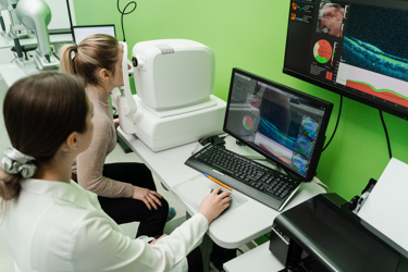
An OCT scan takes only a few minutes and usually requires no special preparation. Sometimes eye drops are used to dilate the pupils beforehand. During the test, the patient rests their chin on a support and focuses on a green target. The scanner captures images one eye at a time, and although a red line may be visible during the process, the scan is painless and does not touch the eye. Remaining still for the minute or two that the scan requires helps ensure clear results.
OCT itself has no risks and causes no discomfort. If pupil dilation is needed, temporary side effects can include blurred vision, sensitivity to light, or a mild headache. These effects wear off within a few hours, but some people arrange for transportation after the appointment since driving may be difficult until vision returns to normal.
The images generated by OCT are reviewed by the provider, who may compare them with earlier scans to track changes. These comparisons can confirm the presence of conditions affecting the retina or optic nerve and provide insight into how advanced they may be. The findings guide decisions about whether to begin treatment, adjust current therapies, or continue monitoring.
In short, OCT is a fast, non-invasive way to see beneath the surface of the eye. By generating detailed, three-dimensional images of retinal and optic nerve layers, it provides critical information that can help specialists diagnose, monitor, and treat a wide range of vision-threatening conditions.
OCT’s Popularity Is Helping Drive Innovation
The expanded use of OCT is driving significant innovation within photonics, particularly in areas such as light-source design, system stability, and image quality validation. One major impact arises from the need for light sources that offer expansive spectral bandwidth combined with exceptionally low noise.
According to Metrology News, a recent industrial breakthrough illustrates this: SuperLight Photonics has developed the SLP-1050, a compact, Class IIIb light source delivering an axial resolution of approximately 2.75 µm and a spectral range extending into the extended short-wave infrared (1,700–2,500 nm), while boasting noise levels three orders of magnitude lower than conventional supercontinuum sources, dramatically enhancing imaging contrast and precision in OCT systems used for industrial inspection of layered and composite materials.
Advances are also emerging on the integrated photonics front, especially in terms of calibration and nonlinearity compensation, writes Optica. A recent demonstration from Fudan University presents a compact, on-chip optical delay line architecture designed for swept-source applications. This spiral silicon nitride waveguide, just 0.8 m in length, achieves a total delay of 10.46 ns with low loss (0.083 dB/cm for TE mode) and, importantly, enables nonlinearity calibration of frequency-swept lasers, which are critical in maintaining depth accuracy in OCT systems.
Another important area of progress is the push to enhance resolution through a combination of hardware refinements and computational techniques. According to arXiv, deep learning–based super-resolution approaches are showing particular promise in addressing the inherent resolution limits of OCT. A recent 2025 study introduced a diffusion model–based plug and play framework that reconstructs high-quality OCT images from sparsely sampled data, producing sharper structural details and improved noise suppression compared to conventional methods. This work demonstrates how computational pipelines can extend imaging performance beyond traditional optical constraints and integrate seamlessly into OCT workflows.
Recent theoretical advances are deepening the understanding of OCT image formation and enabling more precise modeling of optical behavior. A particularly notable contribution is a 2025 study by researchers affiliated with Nikon Corporation and the University of Tsukuba, which formulates a rigorous, four-dimensional image formation theory for OCT and optical coherence microscopy, arXiv writes.
This framework treats the broadband light source using a four-dimensional pupil function – adding light frequency (reciprocal wavelength) as an explicit axis – thus requiring a four-dimensional space–time representation linked to frequency space through a 4D Fourier transform. The result is a highly accurate model that predicts how resolution, aberrations, and dispersion interact in high numerical aperture systems, without relying on simplifying assumptions.
Together, these innovations support a virtuous cycle: enhanced light sources with broader bandwidth and lower noise enable finer spectral calibration, while integrated photonic delay lines help enforce interferometric stability and depth fidelity. Computational approaches and sophisticated imaging theory further permit validation and optimization of depth measurements and resolution across the imaging field. The result is OCT systems delivering more accurate, dependable, and higher-quality imaging, even at greater tissue depths or in challenging industrial environments.
The broader acceptance of OCT is pushing the frontier in photonics across multiple axes: source engineering, on-chip calibration, computational reconstruction, and theoretical modeling. Each advance not only improves OCT performance but also reinforces the development of more compact, robust, and high-precision photonic systems for both biomedical and industrial applications.
