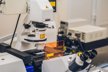
How Hexapods Improve Confocal Microscopy

By John Oncea, Editor

Hexapods enhance confocal microscopy with precise 3D positioning, vibration damping, autofocusing, image stabilization, and large-scale scanning. They're ideal for complex imaging tasks in biological and materials science research, easily integrated with other imaging modalities, and user-friendly.
Hexapod* positioning systems, also known as Stewart Platforms**, are known for their precise and versatile positioning of optical components or instruments. “Hexapods provide 6 degrees of freedom and a user-programmable center of rotation (pivot point) that allows extraordinary flexibility in alignment applications,” writes our friends at PI (Physik Instrumente) LP.
Hexapods are commonly used in photonics and optics for various applications, including optics alignment, fiber optics and silicon photonics alignment, beam steering, satellite antenna testing, high dynamics motion simulation, gyroscopic testing and motion simulation, automated camera image quality testing, computer tomography and 6-axis patient positioning for radiotherapy, automotive and aerospace panel alignment and assembly, and metrology.
Here, we take a look at how hexapods are being used to precisely position and adjust the focus of microscope objectives, sample stages, and other optical components in confocal.
* Hexapods are Parallel Kinematic Manipulators (PKMs)*** that consist of a platform (often a platform with six legs or struts) mounted on a fixed base. Each leg is typically actuated by a linear or rotary motor, allowing for precise control over the position and orientation of the platform.
** “The device that would become the Stewart Platform was first designed by V. Eric Gough of the United Kingdom in 1954,” writes EasyTechJunkie. “Gough was working as an engineer at the Dunlop Tyres Factory in Birmingham, England when he built an early version of the Stewart Platform to be used in the testing of tires. Just over a decade later, D. Stewart presented a paper to the U.K. Institution of Mechanical Engineers proposing an adjustable platform to be used in flight simulators. This is how the device got its name, though some engineers refer to it as a Gough-Stewart Platform in deference to its original inventor.”
*** PKMs, also known as Parallel Robots or Parallel Kinematic Machines, are robotic devices designed as a solution for the limitations presented in Serial Manipulators in terms of stiffness and precision at high accelerations, according to Science Direct.
Confocal Microscopy 101
Confocal microscopy is a specialized form of fluorescence microscopy**** that uses lasers and fluorescence to create a three-dimensional image of a sample, notes Labcompare. A focused laser beam excites molecules at one point of the sample, causing them to release photons and fluoresce. Other key features and principles of confocal microscopy, in bulleted form, include:
- Optical Sectioning: Confocal microscopy uses a pinhole aperture to eliminate out-of-focus light from reaching the detector. This process is called optical sectioning and allows the microscope to capture images from a specific focal plane within the specimen. This results in sharper images with improved contrast and reduced background noise.
- Laser Illumination: Confocal microscopes typically use laser light sources, which produce a focused, intense beam of light at specific wavelengths. These lasers provide high-intensity illumination for the specimen, enabling precise imaging and minimizing photobleaching (damage to the specimen due to excessive light exposure).
- Point Scanning: In confocal microscopy, a single point of the specimen is illuminated and observed at a time. This point is rapidly scanned across the specimen in a predefined pattern. The emitted fluorescence or reflected light from each point is collected and used to construct the final image.
- Fluorescence Imaging: Confocal microscopy is commonly used for fluorescence imaging. Fluorescent labels or dyes are used to mark specific structures or molecules within the specimen. When illuminated with the appropriate wavelength of light (usually from a laser), these labels emit fluorescent light, which is detected and used to create the image.
- Three-Dimensional Imaging: By scanning through different focal planes within the specimen, confocal microscopes can capture multiple optical sections. These sections are then reconstructed to create a three-dimensional representation of the specimen, which is valuable for understanding its structure and organization.
Though it does have slower imaging speeds and comes with higher costs and more complex equipment compared to widefield microscopy, confocal microscopy has proved its worth by helping us better understand cellular and subcellular structures. Offering improved resolution and contrast compared to widefield microscopy, precise optical sectioning for 3D reconstruction, reduction of out-of-focus blur, and minimized photobleaching and phototoxicity due to laser illumination, confocal microscopy has become an indispensable tool in biology, materials science, medicine, and more.
**** In fluorescence microscopy, the entire specimen is flooded with light from a light source, notes Science Direct. In confocal microscopy, only some points of the specimen are exposed to light.
How Hexapods Help Confocal Microscopy Accomplish More
Hexapods play a crucial role in enhancing the capabilities of confocal microscopy by addressing several key challenges and limitations associated with traditional microscopy setups. They are capable of offering highly accurate and stable 3D positioning of the microscope objective or the sample. This precision is of utmost importance for obtaining sharp, clear, and undistorted images, particularly when imaging thick specimens or conducting multi-dimensional scans. The hexapod's capability to precisely manage the position of the sample or objective lens guarantees that the confocal microscope can capture intricate details from different depths within the specimen.
Vibrations from external sources can negatively impact confocal microscopy images. Hexapods dampen vibrations and isolate the microscope, resulting in improved image quality and sharper images. They also can be programmed to automatically adjust the microscope’s focus in real-time, ensuring that the region of interest remains in focus throughout the imaging process. This feature is especially valuable for time-lapse experiments and when studying fast-moving biological processes.
Hexapods enable large-scale sample scanning with sub-micron precision which is useful for imaging entire tissues, organs, or large material samples. Researchers can create high-resolution mosaic images by stitching together multiple confocal microscopy images, all thanks to the hexapod’s precise control over the positioning of the objective or sample.
Confocal microscopy often involves capturing a series of images at different focal planes (Z-stacks) to reconstruct 3D structures. Hexapods make it easier to automate the acquisition of Z-stack images with precise step sizes, ensuring that all relevant information within the specimen is captured accurately.
Hexapods are typically controlled through user-friendly software interfaces that allow researchers to define complex motion paths and scan patterns and can be integrated with other imaging modalities, such as fluorescence microscopy, super-resolution microscopy, and atomic force microscopy, to provide multi-modal imaging capabilities. This allows researchers to combine different imaging techniques to obtain comprehensive information about their samples.
