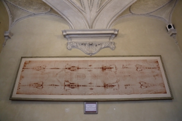
From VP-8 To AI: Five Decades Of Examining The Shroud Of Turin

By John Oncea, Editor

Recent photonic breakthroughs using excimer lasers, X-ray scattering, multispectral imaging, and AI reconstruction are revolutionizing Shroud of Turin authentication research.
Since it was first documented in the 1350s, theologians, historians, and scientists have debated the claim that the Shroud of Turin, a length of linen cloth that bears a faint image of the front and back of a naked man, was Jesus’s shroud upon which his image was miraculously imprinted.
Around 1389, 35 years after it began to be exhibited in the new collegiate church of Lirey, a village in north-central France, the shroud was denounced as a forgery by Bishop Pierre d’Arcis of Troyes, who, in a letter to Antipope Clement VII, reported an artist had confessed to forging the shroud.
The next year, the Avignon antipope Clement VII decreed that, while he would allow the exposition, to “say aloud, to put an end to all fraud, that the aforementioned representation is not the true Shroud of Our Lord Jesus Christ, but a painting or panel made to represent or imitate the Shroud,” according to Time.
“However,” writes Britannica, “subsequent popes, from Julius II on, took the shroud’s authenticity for granted. In 1453, Geoffroi de Charnay’s granddaughter Marguerite gave the shroud to the house of Savoy at Chambéry, and there it was damaged by fire and water in 1532.
The shroud was moved to the royal chapel of the Cathedral of San Giovanni Battista in Turin, Italy, in 1578 and has been preserved there ever since. It has been publicly exhibited only rarely, as on the marriage of Prince Umberto (1931) and on the 400th anniversary of its arrival in Turin (1978).
Religious Leaders Remain Divided
Despite Clement VII’s declaration nearly 700 years ago that the shroud was a fake, debate has raged as to the shroud’s authenticity. One point in favor of the Shroud of Turin’s authenticity is the presence of a unique image with high detail and the cloth’s material consistency with first-century textiles.
Proponents argue that the image’s realistic detail, such as the placement of nail wounds through the wrists rather than the palms, is inconsistent with the knowledge and artistic ability of medieval forgers. Others include:
- The cloth is ancient: The linen textile itself is consistent with fabrics from first-century Israel, a detail that a medieval forger would have had to replicate with great historical accuracy.
- Bloodstains are consistent: Proponents point to the composition of the bloodstains, which include hemoglobin and serum albumin, as being consistent with a real body and not paint.
- Potential historical evidence: Some scholars note historical records of a similar cloth, known as the “Edessa Shroud,” that existed before the 14th century and could be the same relic.
However, starting in the 20th century, people on both sides of the debate began to bolster their arguments with scientific studies. The most significant piece of evidence against the shroud’s authenticity was radiocarbon dating, which estimated the creation of the shroud to have occurred between 1260 and 1390 AD. Opponents argue that the samples were taken from a part of the cloth that had been repaired, making the results inaccurate.
In addition, some forensic studies have concluded that the bloodstains on the shroud are inconsistent with each other and with the image itself, suggesting it is a forgery. And while not a result of scientific study, doubters point out the image bears a resemblance to 14th-century European art, particularly in the face and body, which some scholars believe indicates the shroud is a product of that era.
Analyses Of Authenticity
The Shroud of Turin represents one of archaeology’s most compelling mysteries, and for decades, researchers have employed increasingly sophisticated photonic technologies to decode its secrets.
The modern scientific investigation of the shroud began in 1976 when physicists John Jackson and Eric Jumper analyzed the cloth using the VP-8 Image Analyzer, according to Dr. Richard Kent. This groundbreaking device, originally designed for aerospace applications, converts brightness variations into three-dimensional topographic information.
When applied to ordinary photographs or paintings, the VP-8 produces distorted, meaningless images. However, the shroud yielded something extraordinary: a perfect, anatomically accurate, three-dimensional representation of a human body.
Pete Schumacher, inventor of the VP-8, recalled his astonishment, writing, “I had never heard of the Shroud of Turin before that moment. I had no idea what I was looking at. However, the results are unlike anything I have processed through the VP-8 Analyzer, before or since.”
This unique property indicated that the shroud’s image intensity correlated directly with cloth-to-body distance, encoding spatial information impossible to achieve through conventional artistic techniques.
The STURP Photonic Investigation
The discovery of three-dimensional properties led to the formation of the Shroud of Turin Research Project (STURP), which conducted the most comprehensive scientific examination of the cloth in 1978. Over 120 continuous hours, STURP employed an unprecedented array of photonic and spectroscopic technologies, according to The American Society for Nondestructive Testing.
Optical microscopy up to 1000× magnification revealed the image’s remarkable superficiality, affecting only the outermost fibrils of individual threads. UV spectrophotometry mapped fluorescence patterns across the cloth, while X-ray fluorescence spectrometry identified elemental compositions. Multispectral photography captured images across various wavelengths, revealing details invisible to conventional photography.
Laser-microprobe Raman spectroscopy analyzed molecular structures at microscopic scales, confirming that the image resulted from oxidative degradation rather than applied pigments. STURP’s conclusion was definitive: The image is not the product of an artist. The blood stains are composed of hemoglobin and also give a positive test for serum albumin.
Excimer Laser Breakthrough Research
The most significant recent advance comes from Italy’s ENEA research center, where scientists successfully reproduced shroud-like characteristics using ArF excimer laser technology. Paolo Di Lazzaro’s team employed 193-nanometer deep ultraviolet radiation to achieve unprecedented results. Their excimer laser experiments created permanent yellowing of linen fibers with sub-micrometer depth penetration, exactly matching the shroud’s oxidation pattern.
The ENEA research revealed that achieving shroud-like coloration requires “ultraviolet light necessary to [simulate the image] exceeds the maximum power released by all ultraviolet light sources available today.” According to Renew, the process demands “pulses having durations shorter than one forty-billionth of a second, and intensities on the order of several billion watts,” representing energy levels far beyond medieval technological capabilities.
Dr. Di Lazzaro’s team also documented latent coloration effects, where initially invisible modifications appear only after extended aging, potentially explaining the shroud’s gradual image development over centuries. These findings suggest a two-phase formation process involving initial molecular modification followed by gradual oxidative manifestation.
Advanced X-Ray Analysis Methods
Recent X-ray technologies have revolutionized shroud dating methodology. Dr. Liberato De Caro’s team employed Wide-Angle X-ray Scattering (WAXS) to analyze cellulose degradation patterns in the linen fibers. This non-destructive technique assesses natural aging by measuring crystalline structure deterioration over time.
The WAXS analysis, according to Consiglio Nazionale delle Ricerche, compared the shroud’s cellulose degradation with authenticated first-century samples from Masada, dated between 55-74 AD. Results indicated the shroud’s age as approximately 2,000 years, contradicting the controversial 1988 radiocarbon dating that placed it in medieval times. This discrepancy has prompted extensive reanalysis of the original carbon-14 data, with researchers identifying significant heterogeneity in the samples evaluated.
Multispectral And Hyperspectral Imaging
Contemporary research employs sophisticated multispectral imaging systems operating across ultraviolet, visible, and near-infrared spectra. These technologies, according to the American Society for Nondestructive Testing, reveal previously invisible details while preserving the cloth’s integrity. Hyperspectral analysis provides detailed spectroscopic information for each image pixel, enabling precise chemical mapping without physical sampling.
Recent applications include fluorescence spectroscopy under controlled UV illumination, revealing complex patterns of fiber modification invisible under normal lighting conditions. These techniques have identified microscopic bloodstain characteristics consistent with genuine hemoglobin degradation products rather than artistic materials, according to SciencePG.
Artificial Intelligence Reconstruction
The latest frontier involves artificial intelligence analysis of shroud imagery. According to Academia, advanced algorithms process decades of photonic data to create unprecedented facial reconstructions. AI systems trained on archaeological and forensic datasets have generated detailed three-dimensional models that resolve anatomical features obscured by linen folding and aging effects.
These machine learning applications integrate photogrammetric data with spectroscopic information, producing anatomically consistent reconstructions that maintain coherence across multiple imaging modalities. The AI analysis strengthens forensic evidence supporting the image’s coherent anatomical integrity.
Corona Discharge And Electromagnetic Theories
Giulio Fanti’s research at the University of Padua proposes electrostatic corona discharge as the image formation mechanism. This theory suggests that electromagnetic fields generate brief, intense radiation bursts, creating the observed oxidation patterns. Fourier-transform infrared spectroscopy (FTIR) and Raman spectroscopy support this hypothesis by identifying molecular modifications consistent with electrical discharge effects.
The corona discharge theory explains the image’s vertical directionality and superficial penetration while accounting for the preservation of underlying blood evidence. This mechanism requires no external energy source beyond natural electromagnetic phenomena potentially associated with seismic activity or other geological processes.
Future Photonic Applications
Emerging technologies promise even greater analytical precision. Atom probe tomography offers atomic-level analysis of fiber modifications, while nuclear activation analysis provides ultra-sensitive elemental detection. Terahertz imaging penetrates linen fibers without damage, revealing internal structures invisible to conventional methods.
Quantum-enhanced spectroscopy technologies under development could identify molecular modifications with unprecedented sensitivity, potentially resolving remaining questions about image formation mechanisms. These advances represent the convergence of quantum optics with archaeological analysis.
Scientific Implications
The photonic investigation of the Shroud of Turin demonstrates the remarkable evolution of analytical capabilities over five decades. From the initial VP-8 analysis revealing three-dimensional encoding to recent excimer laser experiments reproducing shroud-like characteristics, these technologies have progressively unveiled the cloth’s extraordinary properties.
