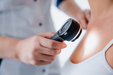
Exploring Non-Invasive Ways To Check For Skin Cancer

By John Oncea, Editor

Two people die of skin cancer in the U.S. every two hours. But, with early diagnosis and treatment, most cases are curable. Now early detection is being made easier with non-invasive methods of checking.
Jimmy Buffet was born on Christmas Day in 1946. Beloved by millions of parrotheads, the man behind such hits as “Margaritaville” and “Cheeseburger in Paradise” combined country, rock, folk, calypso, and pop music with coastal as well as tropical lyrical themes for a sound sometimes called “gulf and western” or tropical rock. Buffet himself referred to his music as “drunken Caribbean rock ‘n’ roll.”
Buffet enjoyed life, dabbling in environmental conservation, charity work, providing disaster relief, and flying planes. He owned a Dassault Falcon 900 jet that he often used while on concert tours and, in August 1994, crashed his Grumman G-44 Widgeon into the waters off Nantucket, MA while attempting to take off.
He was ejected from an NBA game in 2001 for swearing and was detained by French customs officials in Saint Tropez in 2006 for allegedly carrying over 100 pills of ecstasy. Heck, in 2023 a species of crustacean, Gnathia jimmybuffetti, was named after him!
To say Buffet lived a full life would be an understatement, even if you only considered the joy his music brought to his fans. But even in death – he died September 21, 2023 – Buffet created a surge of interest beyond that set aside for the passing of a celebrity.
You see, Buffet died of Merkel cell carcinoma (MCC), a rare and aggressive type of skin cancer that affects the top layer of skin, known as the epidermis. MCC is about three to five times more likely to be deadly than melanoma, and is 40 times rarer than melanoma, with about 3,000 new cases diagnosed in the U.S. every year.
In the days following Buffett’s passing, public interest in MCC skyrocketed, and awareness about this relatively unknown disease was at an all-time high. The Skin Cancer Foundation’s website experienced a steep traffic spike to pages containing information about MCC. This increased awareness is important because Merkel cell carcinoma, while dangerous, is treatable when identified at an early stage.
What Is Skin Cancer?
The Skin Cancer Foundation’s mission is to empower people to take a proactive approach to daily sun protection and the early detection and treatment of skin cancer. To accomplish this, it provides the tools needed to prevent, detect, and treat skin cancer.
The organization’s most crucial role is to help people understand the risks of skin cancer, show what can be done to avoid the disease, and teach people how to spot potential skin cancers at an early stage when they are usually curable.
All of this takes on heightened importance with the understanding that more people are diagnosed with skin cancer in the U.S. each year than all other cancers combined, according to the Skin Cancer Foundation. Equally alarming is the fact that two people die of skin cancer in the U.S. every hour.
The good news is that most cases are curable if they are diagnosed and treated early enough. For instance, the five-year survival rate for melanoma – a less common but more likely to spread type of skin cancer – is 99 percent when detected early.
Skin cancer — the abnormal growth of skin cells — most often develops on skin exposed to the sun. But this common form of cancer also can occur on areas of your skin not ordinarily exposed to sunlight. Skin cancer is often categorized into either melanoma or non-melanoma and non-melanoma skin cancer occurs in either basal or squamous cells:
- Melanoma: This type of cancer is less common than the other types, but it's more likely to spread to other parts of the body.
- Basal cell carcinoma (BCC): These cancers often develop on the head, neck, and arms after years of sun exposure or tanning. BCCs can grow deep and form anywhere on the body, including the chest, abdomen, and legs.
- Squamous cell carcinoma (SCC): These cancers generally grow faster than BCCs and start in keratinocytes found in the epidermis. Most SCCs develop on skin exposed to the sun.
As alluded to a couple of times already, early detection saves lives. Learning what to look for on your skin gives you the power to detect cancer early when it’s easiest to cure before it can become dangerous, disfiguring, or deadly.
Non-Invasive Optical Methods For Detecting And Treating Skin Cancer
“Historically, the diagnosis of skin cancers has depended on various conventional techniques which are of an invasive manner, writes the National Center for Biotechnology Information (NCBI). In recent years, however, non-invasive skin cancer diagnostic methods have been used to detect skin cancer.”
These include photography, dermoscopy, sonography, confocal microscopy, fluorescence spectroscopy, optical coherence tomography, the multispectral imaging technique, thermography, electrical bio-impedance, tape stripping, and computer-aided analysis.
Raman spectroscopy, a technique that is used to discover various modes in a system that involves vibrational, rotational, and other low–frequency modes, is also used. “It depends on Raman scattering of monochromatic radiations, usually from a laser in the visible, near-infrared, and near UV rays,” NCBI writes. “In Raman scattering, inelastic collisions take place between the photons of an irradiating laser beam and the sample (or tissue) molecules. The obtained spectra can be processed and analyzed to provide automated feedback at the time of measurement. This system provides better sensitivity in differentiating the tissues.”
Last month, according to Interesting Engineering, advancements were made by Queen Mary University of London and the University of Glasgow regarding the use of terahertz (THz) spectroscopy, an imaging technique that is used to detect epithelial cancers.
The newly developed biosensor has a high sensitivity for detecting skin cancer compared to other, often time-consuming approaches and leverages THz waves to provide a non-invasive method for analyzing underlying tissue properties.
“Traditional methods for detecting skin cancer often involve expensive, time-consuming, CT, PET scans and invasive higher frequencies technologies,” explained Shohreh Nourinovin, postdoctoral research associate at Queen Mary, and the study’s first author. “Our biosensor offers a non-invasive and highly efficient solution, leveraging the unique properties of THz waves – a type of radiation with lower energy than X-rays, thus safe for humans – to detect subtle changes in cell characteristics.”
The biosensor’s design holds the key innovation, featuring tiny, asymmetric resonators on a flexible substrate that can pick up on subtle changes in cell properties. Unlike traditional methods that rely solely on refractive index, this device analyses a combination of parameters, including resonance frequency, transmission magnitude, and a value called “Full Width at Half Maximum” (FWHM).
This comprehensive approach provides a richer picture of the tissue, allowing for more accurate differentiation between healthy and cancerous cells and measuring the degree of malignancy in the tissue. In tests, the biosensor successfully differentiated between normal skin cells and basal cell carcinoma (BCC) cells, even at different concentrations.
“The implications of this study extend far beyond skin cancer detection,” says Nourinovin. “This technology could be used for early detection of various cancers and other diseases, like Alzheimer’s, with potential applications in resource-limited settings due to its portability and affordability.”
There’s also millimeter-wave imaging, a non-invasive cancer-detecting method that uses the same technology as an airport security scanner. According to ScienceDaily, millimeter-wave rays can penetrate up to 2 millimeters into human skin, creating a 3D map of scanned skin lesions, and detecting changes in tissue layers of excised organs or different skin layers where most skin tumors originate.
In an active scanner, two antennas transmit millimeter waves simultaneously as they rotate around the body. The wave energy reflected from the body or other objects on the body is used to construct a three-dimensional image, which is displayed on a remote monitor for analysis. The captured signals are then combined and processed to form the image of the target.
Millimeter-wave imaging can produce high-contrast, high-resolution 3D images of the skin, which identify the locations of the tumors as accurately as histological imaging. The technology has the potential to vastly reduce or even eliminate unnecessary biopsies by quickly identifying malignant and benign tissues.
A large-scale study showed that there are statistically significant contrasts between the millimeter-wave dielectric properties of normal skin and two of the most common types of skin cancer, basal cell carcinoma (BCC) and squamous cell carcinoma (SCC).
BONUS: Adding Big Data To The Mix
The increasing use of non-invasive methods to evaluate and diagnose skin cancer is creating more and more data. So much so that national cancer registries are only able to track a small proportion of skin cancers, according to University of Arizona Health Sciences.
“Basal cell and squamous cell carcinomas, which are both non-melanoma skin cancers, account for 97% of skin cancers diagnosed in the U.S. and are not required to be reported to state cancer registries. To gain a more complete picture of skin cancer and address gaps in diagnostic and prognostic tools, University of Arizona Health Sciences researchers created a database for melanoma and non-melanoma skin cancer that links patient data, images, and tissue.
Researchers can use the Patient Registry, Imaging Database, and Tissue Bank (PRIT) to develop innovative tools that can assist doctors in diagnosing and predicting skin cancer outcomes. This resource can help in developing artificial intelligence software that can recognize, diagnose, and classify skin cancers based on the risk of metastatic disease. It also can aid in discovering biomarkers and identifying new targets for effective preventive and therapeutic strategies.
“PRIT is greater than the sum of its parts, but you can think of it as being built on three pillars,” said Nirav Merchant, director of the university’s Data Science Institute and interim director of the Center for Biomedical Informatics and Biostatistics at UArizona Health Sciences. “It brings together three separate computer programs to make them more manageable and clinically useful. This allows Dr. Curiel-Lewandrowski’s team and potentially other researchers, centers, and universities to field a larger body of data sets and make decisions in a consistent and organized manner.”
PRIT is a system that brings together three programs - REDCap, OpenSpecimen, and OMERO. REDCap is a web application that allows researchers to create and manage online surveys and databases securely. OpenSpecimen is a bio-banking platform that helps to collect, store, process, annotate, and distribute biospecimens for research purposes. OMERO is a software that enables researchers to manage, visualize, and analyze microscopy images and their associated metadata.
By integrating these three platforms, researchers can now access a comprehensive database that contains anonymized patient data, including information such as age, gender, and location, as well as a bank of biospecimens and a library of skin cancer images. All of this information is now accessible from one central location and linked together, making it easier for researchers to analyze and draw insights from the data.
“PRIT is a great example of how investing in technology development can have unforeseen benefits,” Merchant said. “The next steps would be to bring in more modern machine learning and AI techniques. All these images are now labeled and connected to libraries of other data. When you bring in an unknown image, AI could pull up the closest matching image and provide a repository of other information about it. It is hard for a human to look at an image of skin cancer and describe it in nuanced detail that would separate it from all the other images of skin cancer. But computers are very good at sorting and matching images.”
