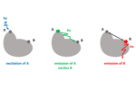
Total internal reflection fluorescence (TIRF) microscopy is used to watch biological processes unfold in real time. Taking advantage of the ability to label individual molecules with different colors of fluorescent tags, TIRF microscopy lets scientists view the complex molecular assemblies that govern cellular processes.
Complex ratiometric measurements are dependent on many factors — encompassing chemistry, instrumentation, and analysis choices — that can affect sensitivity and, by extension, the ability to discern meaningful quantitative information from the data.
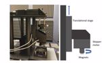
Magnetic tweezer experiments often seek to measure changes in the extension or relaxation of a polymer, a functionality useful, for example, in exploring how different enzymes manipulate polymer structures.
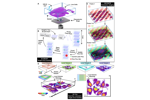
Single-molecule microscopy techniques facilitate direct study of molecular mechanisms, enabling leaps in understanding how viruses assemble, disassemble, and interact with their hosts.
A team of Duke University researchers sought to explore the viral infection process before the virus has bonded to tissue, understanding how viruses navigate the epithelial space. To do so, they needed a microscope capable of observing that journey in a tissue culture model that closely replicates the lungs.
The RM21 microscope is suitable for a variety of microscopy and nanoscopy methods. Each of the four standard models has been engineered for precision alignment and stability for advanced microscopy.