UK Scientists Invent Camera That Can 'See Through' Human Body
By Jof Enriquez,
Follow me on Twitter @jofenriq

Scientists at the University of Edinburgh and Heriot-Watt University, through the Proteus Interdisciplinary Research Collaboration, have developed a new camera that can image individual photons emanating from fiber optic medical instruments inside the human body.
Common medical procedures, such as endoscopy, typically rely on X-rays to confirm the correct placement of an invasive probe as it enters and moves through the digestive track. X-rays, however disrupt the continuity of the procedure, and carry exposure risks to the patient.
Other scientists have proposed having patients swallow tiny pill-sized cameras, using optical filters, or attaching microendoscopes to conventional endoscopes, to conduct better imaging and biopsies.
Now, a research group at the University of Edinburgh and Heriot-Watt University is proposing a non-invasive alternative: a small external camera that is able to detect the illuminated tip of an endoscope, even though the instrument emits photons that tend to scatter or bounce off tissue, rather than pass through the body.
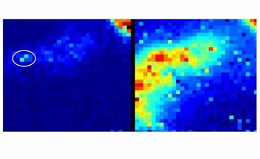
Some photons do penetrate through body tissue, though, and the new imaging device is so sensitive that it is able to capture these "ballistic and snake photons using a time resolved single-photon detector array," according to the study published in Biomedical Optics Express.
The camera's single photon detectors that are integrated onto a silicon chip also are able to detect the scattered light, in addition to the light that passes through tissue and straight onto the camera. In addition, the device can record the time taken for light to pass through the body and work out exactly where the endoscope is located in real time, which is something that we cannot do with x-rays due to the heavy exposure.
"We're overlaying the location of the device we're trying to find, on top of some conventional imaging, so this is more like seeing a glowing spot showing you where the device is," says Dr. Mike Tanner, research fellow at Heriot-Watt University, in a BBC video.
The prototype camera has been shown in experiments to be able to track the location of a point light source through 20 centimeters of tissue under normal light conditions. The device could soon jump from the physics lab into practical and wider use in operating rooms and at the bedside.
"The ability to see a device’s location is crucial for many applications in healthcare, as we move forwards with minimally invasive approaches to treating disease," says Kev Dhaliwal, Professor of Molecular Imaging and Healthcare Technology, University of Edinburgh and Clinical Lead, Proteus.
