Manufacturing A More Accurate Bioimaging Protein
By Chuck Seegert, Ph.D.

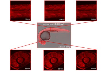
Using certain cells lines, scientists at the Helmholtz Zentrum München research center in Germany have found a way to manufacture the bioimaging protein Amrose. The novel approach provided the cells with the right materials and then let them formulate into the complex molecule independently.
Bioimaging is used to visualize structures and processes in biological organisms including humans. Tumors and metastasis can be tracked, as can the responses to a drug that’s applied. These markers can even be used for whole-body imaging. Bioimaging works because of the marker’s ability to be excited or stimulated by infrared light, which is followed by the emission of light in another wavelength. This secondary emission is then measured by the detection system.
Some markers in the past have been excited by wavelengths of near-infrared light, but the markers emit wavelengths that are relatively close to their excitation wavelength. This can lead to challenges in detection, as the two wavelengths get confused by the detector. According to a recent press release, having a biomarker that could be excited by far-infrared and emit in the infrared spectrum was the goal of the team of scientists from Helmholtz Zentrum München. This type of biomarker would increase the sensitivity of bioimaging.
Most notably, however, was how the team went about synthesizing the protein they call Amrose. Instead of running complex reactions with beakers and chemicals in the lab, they turned to cellular machinery. In a research study published in PLOS one, the researchers provide details to their approach which revolved around the avian B cell line DT40, a form of white blood cell that creates antibodies.
In B-cells, antibodies are produced using a process of protein evolution, according to the study. To develop highly specific antibodies for foreign material (antigens) that may invade the body, the cells use an iterative process until the proteins match closely to the antigen. Using this cellular machinery, the team was able to induce protein production that led to the Amrose molecule they were seeking — that is, a molecule excited in the far infrared wavelengths.
What’s amazing about this is that the researchers had no prior knowledge of what the protein they were looking for would be like from a structural standpoint. According to the study, they let the cells determine what was needed to achieve their goals.
“Here we have demonstrated the further use of this novel technology to develop highly sought after biologically relevant fluorescent markers quickly and easily for different imaging needs,” said Randolph Caldwell, lead researcher of the study, in the press release.
Ways of enhancing biomarkers is an area of ongoing research. Recently, a team form MIT showed that by manipulating the direction of emitted light, biomarkers could be improved for many bioimaging applications.
Image Credit: PLOS one
